

![]()
All of these mole pictures (with exception of the bottom photo) are of biopsy-proven deadly malignant melanoma skin cancers that our clinic has managed for patients. Photos are copyright (c) The Mole Clinic and must not be copied or distributed without express written consent.
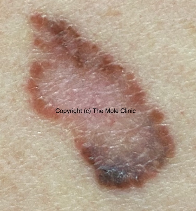 |
A - Asymmetrical ShapeMelanoma lesions are often irregular, or not symmetrical, in shape. Benign moles are usually symmetrical. |
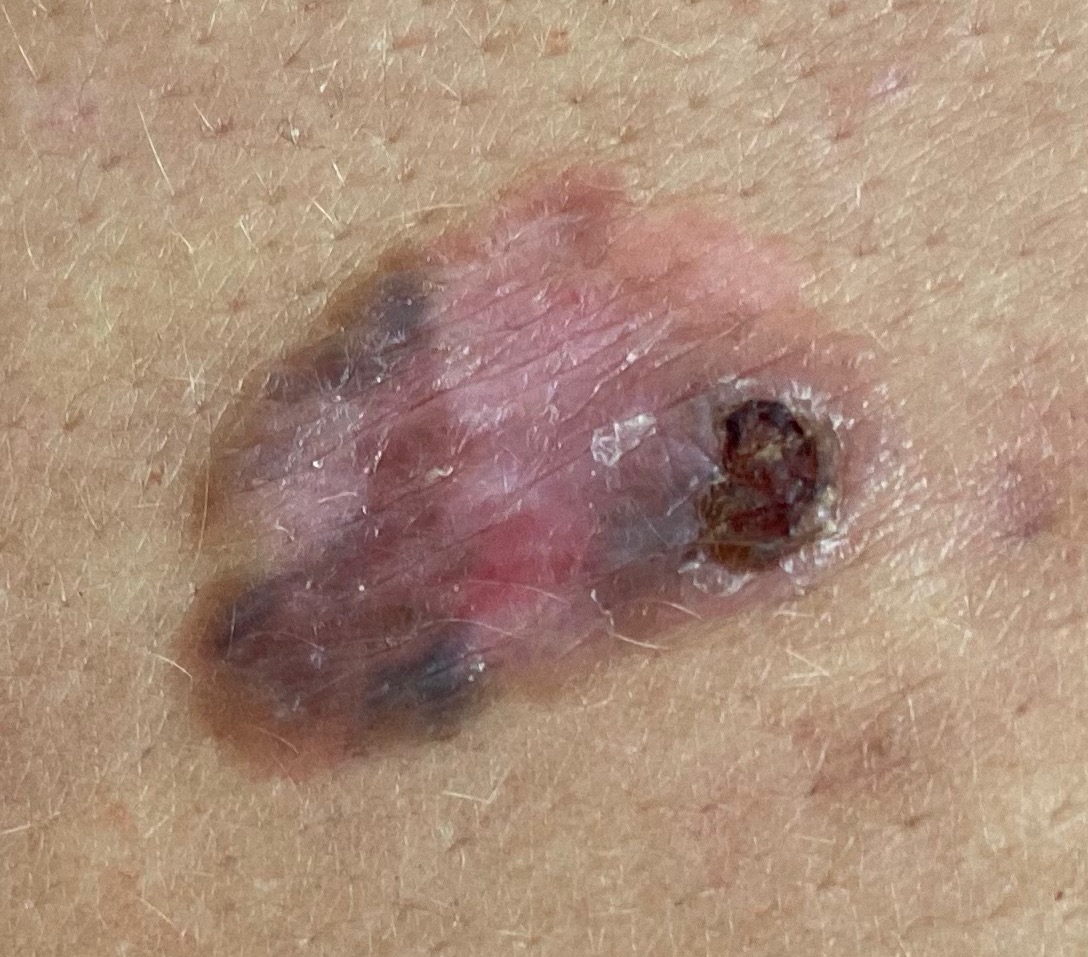 |
B - BorderTypically, non-cancerous moles have smooth, even borders. Melanoma lesions usually have irregular borders that are difficult to define. In this photo, there is also a scab present. This is an area where cancer cells are rapidly dividing outgrowing their blood supply and causing an area of skin breakdown. Moles that itch or bleed are concerning and should be assessed by a medical professional. |
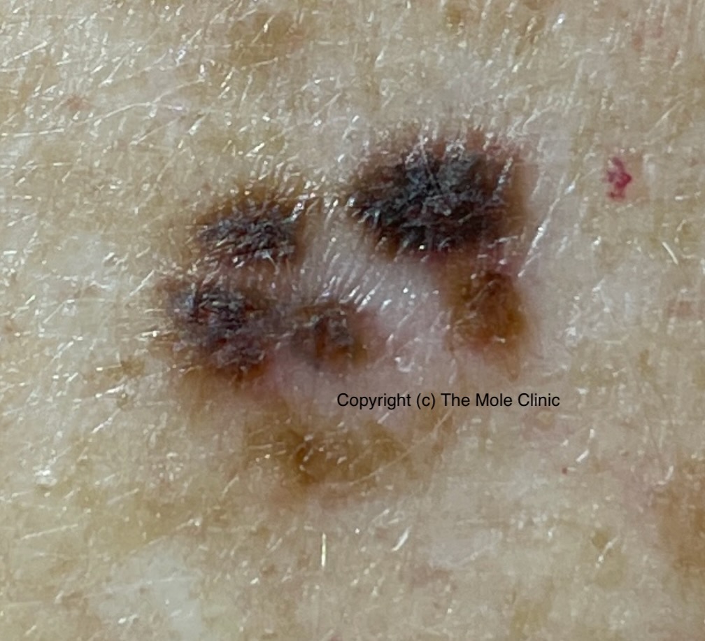 |
C - ColorThe presence of more than one color (blue, black, brown, tan, etc.) or the uneven distribution of color can sometimes be a warning sign of melanoma. Benign moles are usually a single shade of brown or tan. |
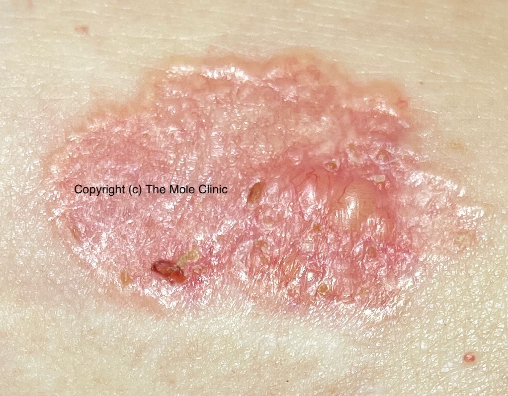 |
D - DiameterMelanoma lesions are often greater than 6 millimeters in diameter (approximately the size of a pencil eraser). The photo on the left was taken of a non-Melanoma skin cancer called Basal Cell Carcinoma. The lesion in this photo was about 2.5cm in maximal dimension and located on a woman's tummy. |
If you have any concerns, please do not hesitate to contact the clinic, or call your family doctor.